
Private Carotid Duplex Ultrasound with CIMT Assessment
A comprehensive Doppler ultrasound assessment of the arteries supplying the brain, designed to evaluate carotid plaque, narrowing (stenosis), blood-flow velocities and carotid artery wall thickness — with carotid intima-media thickness (CIMT) measured where clinically appropriate.
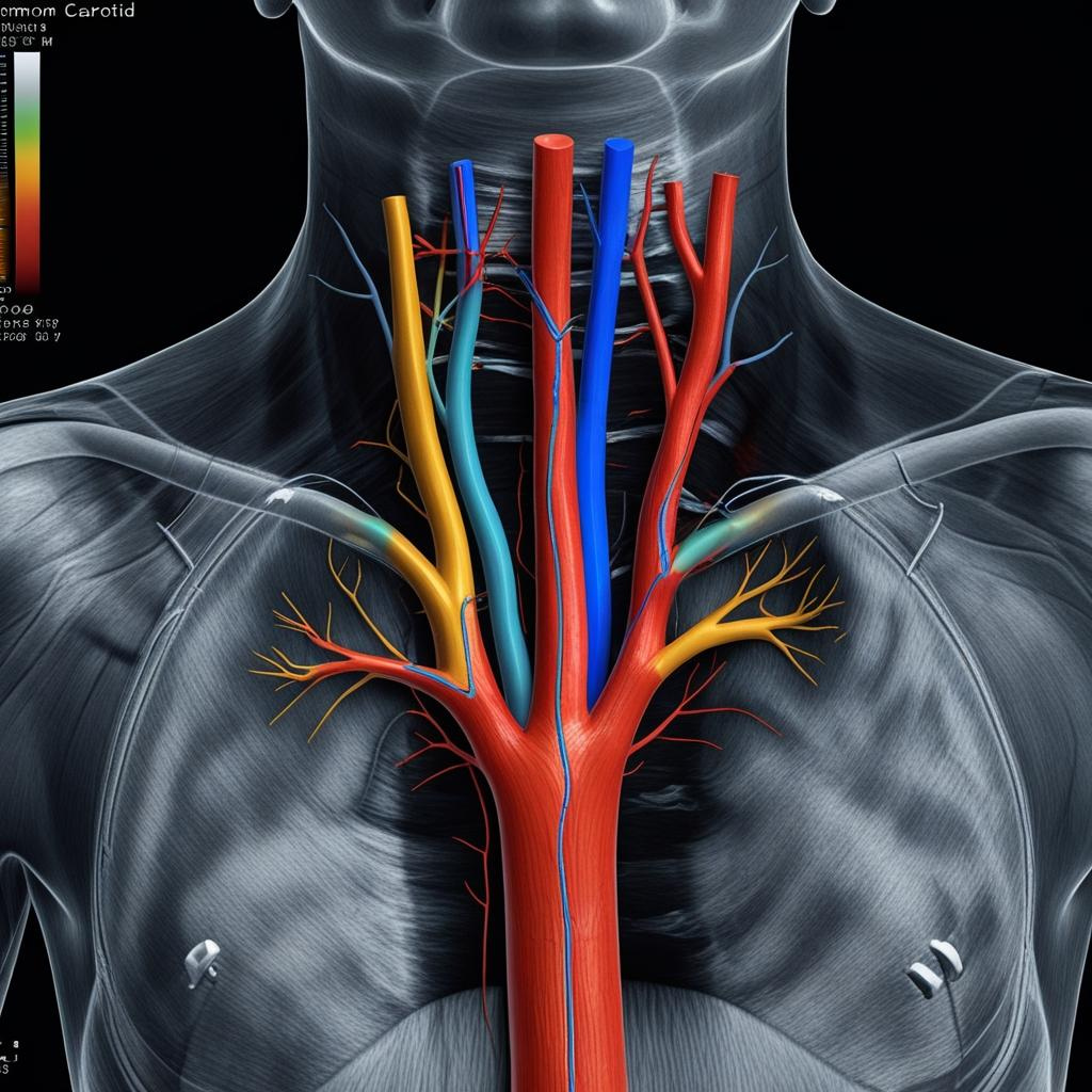
Detailed assessment of the arteries supplying the brain
The carotid arteries run on either side of the neck and provide a major supply of blood to the brain. A carotid duplex ultrasound combines high-resolution structural ultrasound with colour and spectral Doppler to assess the artery wall, identify atherosclerotic plaque and evaluate how blood is flowing through the vessel.
The examination can detect carotid plaque and assess whether narrowing is present. Doppler velocity measurements help determine whether a stenosis is causing significant alteration of blood flow. Vertebral artery flow can also be evaluated as part of the vascular examination.
UKSONO’s carotid duplex examination also includes carotid intima-media thickness (CIMT) measurement where clinically appropriate. CIMT provides information about arterial wall thickness, but it is interpreted alongside plaque findings and established cardiovascular risk factors rather than as a stand-alone diagnosis or direct prediction of an individual’s future stroke or heart attack.
More than simply checking for a blockage
A comprehensive carotid duplex examination evaluates arterial anatomy, plaque burden, stenosis and blood-flow characteristics rather than relying on one measurement alone.
Common carotid arteries
Assessment of the right and left common carotid arteries, including vessel-wall appearance, plaque and Doppler flow.
Internal carotid arteries
The internal carotid arteries are assessed for plaque, narrowing and elevated blood-flow velocities that may indicate significant stenosis.
External carotid arteries
Vessel appearance and Doppler flow are evaluated where relevant as part of the complete carotid examination.
Vertebral artery flow
Direction and character of vertebral artery flow may be assessed to provide information about posterior circulation blood flow.
Atherosclerotic plaque
Where plaque is identified, its location, distribution and relevant ultrasound appearance are documented.
Blood-flow velocities
Peak systolic and end-diastolic velocities are measured using spectral Doppler to support assessment of carotid narrowing.
How ultrasound evaluates both artery structure and blood flow
The term duplex refers to combining anatomical ultrasound imaging with Doppler assessment of moving blood.
B-mode ultrasound
High-resolution greyscale imaging demonstrates the carotid artery wall, lumen and visible atherosclerotic plaque.
Colour Doppler
Colour Doppler demonstrates the direction and pattern of blood flow and can help identify areas of disturbed or accelerated flow.
Spectral Doppler
Spectral Doppler produces measurable blood-flow waveforms and velocities that are used together with imaging findings to assess stenosis.
What is CIMT — and what can it actually tell you?
Carotid intima-media thickness (CIMT) is an ultrasound measurement of the combined thickness of the intimal and medial layers of the carotid artery wall. It is commonly measured within a plaque-free segment of the distal common carotid artery using a high-resolution longitudinal ultrasound image.
CIMT has been extensively studied as a marker of arterial wall change and has associations with cardiovascular risk factors and cardiovascular events at population level.
The Mannheim CIMT consensus emphasises the importance of standardised image acquisition and, importantly, distinguishes diffuse carotid intima-media thickening from focal atherosclerotic plaque.
At UKSONO Healthcare, CIMT is therefore reported in clinical context . A CIMT result is not presented as a stand-alone “stroke-risk score”, and it does not replace established cardiovascular risk assessment including blood pressure, cholesterol, diabetes status, smoking history and clinical medical review.
CIMT measurement concept: the distance between the lumen–intima and media–adventitia interfaces within the carotid artery wall.
The Mannheim consensus defines carotid plaque as a focal structure encroaching into the arterial lumen by at least 0.5 mm, or by at least 50% of the surrounding IMT value, or demonstrating a thickness greater than 1.5 mm.
This is one reason plaque assessment should be considered separately from CIMT measurement.
Why carotid plaque assessment matters
Atherosclerotic plaque can narrow the carotid lumen and alter blood flow. Duplex ultrasound allows the artery wall, plaque and Doppler velocities to be assessed together.
Plaque detection
Ultrasound can directly visualise focal atherosclerotic plaque, particularly around the carotid bifurcation and proximal internal carotid artery.
Stenosis assessment
Vessel appearance, colour Doppler and spectral Doppler velocities are combined to evaluate whether narrowing is haemodynamically significant.
Appropriate clinical follow-up
Significant carotid findings may require medical review through your GP, stroke/TIA service, vascular specialist or another relevant clinical pathway.
Who may consider a carotid duplex ultrasound?
The usefulness of carotid ultrasound depends on the clinical situation. It should not be marketed as indiscriminate population screening for every healthy person.
Cardiovascular risk factors
Selected patients with hypertension, diabetes, dyslipidaemia, smoking history or other vascular risk factors may discuss whether carotid assessment is appropriate.
Known vascular disease
Carotid ultrasound may be clinically relevant where atherosclerotic vascular disease has already been diagnosed.
Carotid bruit
A carotid bruit identified during clinical examination may prompt vascular imaging depending on the wider clinical context.
Previous carotid disease
Follow-up imaging may be recommended where carotid plaque or stenosis has previously been identified.
Cardiovascular risk discussion
Plaque and CIMT information may add vascular context for selected patients, but these findings complement rather than replace standard cardiovascular risk assessment.
Clinician recommendation
A GP, cardiologist, neurologist or vascular clinician may recommend carotid duplex based on symptoms, examination or previous investigations.

A carotid scan is one part of cardiovascular prevention
Carotid ultrasound can demonstrate arterial plaque, stenosis and blood-flow abnormalities, but cardiovascular prevention depends on the wider clinical picture.
Blood pressure, cholesterol, diabetes, smoking status, physical activity, family history and other established risk factors remain important determinants of cardiovascular risk.
Where plaque or increased arterial wall thickness is identified, the result should be considered by an appropriate clinician together with the individual’s overall risk profile.
What happens during a carotid Doppler ultrasound?
The examination is non-invasive, usually painless and requires no needles or radiation.
Clinical history
Your relevant vascular history, medications and reason for the examination are reviewed.
Neck ultrasound
You lie comfortably with your neck gently positioned while both sides are examined using a high-frequency linear ultrasound probe.
Doppler measurements
Colour and spectral Doppler are used to assess flow direction, waveforms and blood-flow velocities.
Results & report
The main findings are explained and a written report documents the examination and recommended next steps.
Comfortable, non-invasive carotid vascular imaging
During a carotid duplex examination, the ultrasound transducer is positioned directly over the carotid arteries on each side of the neck.
High-resolution B-mode imaging is combined with colour Doppler and spectral Doppler to evaluate vessel-wall structure, plaque, blood-flow direction and measured flow velocities.
You will normally lie on your back with the neck gently extended and turned slightly away from the side being examined. Ultrasound gel is applied to the neck and the probe is moved gently along the course of the carotid arteries.
There is no ionising radiation and no injection is normally required.

Carotid duplex ultrasound — £240
A detailed bilateral vascular ultrasound assessment with Doppler measurements and CIMT where clinically appropriate.
Private Carotid Duplex Ultrasound
Comprehensive bilateral carotid Doppler ultrasound assessing plaque, blood flow and carotid stenosis.
What Our Patients Say
Read independent patient feedback from people who have chosen UKSONO Healthcare for diagnostic ultrasound.
Evidence and guidance behind carotid ultrasound and CIMT
For patients and clinicians who want to explore the scientific background, recognised methodology and limitations of CIMT and carotid ultrasound.
Read the full article →
Read the systematic review →
Read NICE guidance →
Read guideline →
Carotid duplex ultrasound FAQs
Answers to common questions about carotid plaque, stenosis, Doppler velocities, CIMT, stroke risk and preparation.
What is a carotid duplex ultrasound?
Which arteries are examined?
What is carotid plaque?
What does carotid stenosis mean?
What is CIMT?
Why measure CIMT?
Does increased CIMT mean I will have a stroke?
Is CIMT the same as carotid plaque?
What thickness counts as carotid plaque?
Should I repeat CIMT every year to see if it improves?
Can this scan tell me my exact stroke risk?
Is carotid ultrasound recommended for everyone as a screening test?
Can carotid ultrasound detect a previous stroke?
What happens if significant carotid stenosis is found?
What if I currently have stroke-like symptoms?
Do I need a GP referral?
Does carotid ultrasound hurt?
Does carotid Doppler ultrasound use radiation?
How should I prepare for the scan?
How long does a carotid duplex scan take?
Will I get my results on the same day?
How much does a private carotid duplex ultrasound cost?
Book your private carotid duplex ultrasound
Select an available appointment through UKSONO Healthcare’s secure online booking system.
UKSONO Healthcare Online Booking
Carotid Duplex Ultrasound · £240
Private carotid ultrasound in Kent & London
Carotid duplex ultrasound appointments are available through UKSONO Healthcare in Tunbridge Wells, Orpington and Canary Wharf.
UKSONO Healthcare
36 Monson Road
Tunbridge Wells
UKSONO Healthcare
Orpington Health & Wellbeing Centre
19 Homefield Rise
Orpington
BR6 0FE
UKSONO Healthcare
30 S Colonnade
Canary Wharf Estate
London
E14 5HX
Need help with your booking?
Call 01892 251532
Book your carotid duplex ultrasound
Comprehensive bilateral assessment of carotid plaque, stenosis, blood-flow velocities and CIMT where clinically appropriate — with clear reporting and onward guidance.