
Private Breast Ultrasound & Axilla Scan
A detailed, sonographer-led breast ultrasound assessment for breast lumps, focal breast pain, cysts, fibroadenoma, axillary concerns, breast implants and other focal breast symptoms — with same-day verbal feedback and a comprehensive written report.
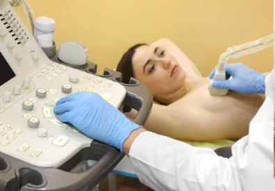
Detailed breast ultrasound assessment with clear answers
Breast ultrasound is a non-invasive imaging examination that uses high-frequency sound waves to assess the internal structure of breast tissue.
It is particularly useful for evaluating a palpable breast lump, focal area of breast pain, cystic or solid lesions, previously identified benign abnormalities, selected nipple or peri-areolar concerns, axillary lymph nodes and tissues surrounding breast implants.
Ultrasound can help determine whether an abnormality is fluid-filled or solid and allows assessment of features including shape, margins, echogenicity, orientation, posterior acoustic behaviour and vascularity where clinically relevant.
Your examination is interpreted in the context of your symptoms and clinical history. Where a finding cannot safely be considered benign, UKSONO Healthcare provides clear advice regarding the appropriate onward pathway.
Reasons patients book a private breast ultrasound
Breast symptoms are common and many causes are benign. A targeted ultrasound can provide additional imaging information, while symptoms that require specialist breast-clinic assessment should still follow the appropriate clinical pathway.
Breast lump or palpable area
Assessment of a focal lump, thickening or palpable area to determine whether an ultrasound abnormality is present and to characterise its imaging features.
Focal breast pain
Targeted assessment of persistent or localised breast pain, particularly where there is a specific area of tenderness or associated palpable concern.
Breast cyst
Ultrasound is particularly useful for identifying fluid-filled breast cysts and determining whether their appearances are simple, complicated or require additional assessment.
Fibroadenoma assessment
Evaluation or follow-up of a previously identified fibroadenoma, taking account of its ultrasound morphology, size and any reported interval change.
Axillary lump or swelling
Assessment of the underarm region and lymph nodes where there is focal swelling, discomfort or a palpable axillary abnormality.
Breast implant concern
Assessment of the peri-implant tissues and selected sonographic features that may be associated with implant-related complications.
Previous breast surgery
Targeted review of a palpable or symptomatic area following previous surgery or intervention, including assessment for fluid collections, scar-related changes or fat necrosis.
Pregnancy or breastfeeding
Ultrasound is commonly used to investigate focal breast symptoms during pregnancy and lactation because it does not use ionising radiation.
Previous benign finding
Follow-up ultrasound may be appropriate for selected previously assessed benign findings where interval imaging has been advised.
A responsible pathway for symptomatic breast assessment
Breast ultrasound can provide valuable information, but safe breast assessment also means recognising when ultrasound should not be the end of the pathway.
We assess the ultrasound finding
The breast and clinically relevant axillary tissues are examined and any focal abnormality is characterised using recognised ultrasound features.
We explain significant findings
If the examination demonstrates an indeterminate or suspicious finding, this is explained clearly and documented in the ultrasound report.
We support onward specialist assessment
With the patient’s consent, clinically significant findings can be communicated to their GP to facilitate appropriate NHS breast-clinic assessment and further investigation.
Mammography is commonly an important component of symptomatic breast imaging from age 40. Ultrasound does not replace mammography, the NHS Breast Screening Programme, clinical breast examination or specialist triple assessment where these are indicated.
Common findings seen on breast ultrasound
Breast ultrasound can demonstrate a wide range of benign, inflammatory and potentially suspicious abnormalities. The imaging appearance determines whether reassurance, surveillance or further specialist investigation is appropriate.
Simple breast cysts
Cysts are fluid-filled structures and are common within breast tissue. A typical simple cyst has characteristic ultrasound appearances including anechoic fluid content, a thin wall and posterior acoustic enhancement.
Fibroadenoma
Fibroadenomas are common benign solid breast lesions, particularly in younger people. Typical lesions may appear oval, well-defined and orientated parallel to the skin.
Fibrocystic change
Hormonal changes can cause breast lumpiness, discomfort and cyst formation. Ultrasound may demonstrate small cysts and other benign changes within the breast tissue.
Duct ectasia and duct changes
Ultrasound can demonstrate dilated ducts and selected abnormalities around the retroareolar region, particularly where there is a focal nipple or peri-areolar concern.
Mastitis and breast abscess
Ultrasound may demonstrate inflammatory tissue change, increased vascularity or a focal fluid collection where mastitis or abscess is clinically suspected.
Fat necrosis
Fat necrosis may follow trauma, surgery or other breast procedures and can sometimes present as a palpable lump. Its appearance can vary and may occasionally require further imaging correlation.
Lymph nodes
Ultrasound assesses lymph-node morphology including cortical thickness, shape and the appearance of the fatty hilum. Indeterminate or suspicious nodes may require specialist review.
Peri-implant abnormalities
Ultrasound can assess peri-implant tissues and may demonstrate fluid collections or sonographic features associated with selected implant complications. MRI may still be required for further assessment in some situations.
Indeterminate or suspicious mass
Irregular shape, non-circumscribed margins, non-parallel orientation, posterior shadowing and other suspicious features can increase concern and require appropriate specialist breast assessment and potentially tissue sampling.
Understanding your U1–U5 breast ultrasound result
UK breast imaging uses a standard numerical classification to communicate the level of imaging concern. For ultrasound, the category is prefixed with the letter U.
Normal
No significant abnormality is identified on ultrasound.
Benign
The imaging finding demonstrates benign ultrasound features.
Indeterminate / probably benign
There is a small likelihood of malignancy and further investigation is indicated.
Suspicious
The imaging appearance is suspicious for malignancy and further investigation is indicated.
Highly suspicious
The imaging features are highly suspicious for malignancy and require urgent specialist assessment.
More than simply scanning the area of concern
The examination is tailored to the clinical indication and incorporates relevant breast and axillary ultrasound assessment.
Relevant clinical history
Discussion of your symptoms, duration, location, relevant family history, previous surgery, breast implants and previous imaging where available.
Targeted breast assessment
Detailed ultrasound of the symptomatic or clinically relevant breast tissue using a high-frequency transducer.
Axillary assessment
Ultrasound assessment of the relevant underarm lymph-node region where clinically indicated.
Doppler evaluation
Colour or power Doppler may be used where clinically appropriate to assess vascularity within or around a finding.
Immediate explanation
Your sonographer explains the main ultrasound findings and answers relevant questions following the examination.
Written ultrasound report
A formal report documents the examination, imaging findings, impression and recommended next steps where required.
What happens during a breast ultrasound?
The procedure is non-invasive, does not use ionising radiation and usually requires no special preparation.
Clinical discussion
We first establish the exact reason for your scan and the area requiring assessment.
Positioning
You will usually lie on the examination couch with the arm raised on the side being examined.
Ultrasound examination
Ultrasound gel is applied and the breast is examined using a high-frequency ultrasound transducer.
Results and next steps
The main findings are explained and your formal report documents any recommendation for further assessment.
Breast ultrasound and mammography are not the same test
Different breast imaging tests answer different clinical questions. One should not automatically be considered a substitute for another.
| Feature | Breast Ultrasound | Mammography |
|---|---|---|
| Technology | High-frequency sound waves | Low-dose X-ray imaging |
| Radiation | No ionising radiation | Uses low-dose ionising radiation |
| Useful for | Characterising focal abnormalities, distinguishing many cystic and solid lesions and assessing selected axillary findings | Broader breast assessment, calcifications and abnormalities that may not be visible on ultrasound |
| Younger symptomatic patients | Commonly used as the initial imaging examination where appropriate | Used according to age, clinical findings and specialist assessment |
| Age 40+ | May provide important complementary information | Commonly an important initial component of symptomatic breast imaging |
| NHS screening replacement? | No | NHS mammography screening follows its own national programme |
Why appropriate breast assessment matters
Most breast symptoms do not automatically mean cancer, but a new or persistent breast change should be assessed appropriately.
Lifetime breast cancer risk
Cancer Research UK estimates that around 1 in 7 women in the UK will develop breast cancer during their lifetime.
Source: Cancer Research UKMany breast lumps are not cancer
Breast lumps are common and many have benign causes, but the NHS advises that a breast lump should still be clinically checked.
Source: NHSIonising radiation
Diagnostic breast ultrasound uses high-frequency sound waves rather than X-rays and therefore does not expose you to ionising radiation.
Private breast ultrasound prices
Choose the breast ultrasound assessment that best reflects your clinical needs.
One Breast Ultrasound
Focused ultrasound assessment of one breast for a unilateral symptom or localised area of concern.
Both Breasts & Axillae
Comprehensive bilateral breast ultrasound with relevant axillary assessment and side-to-side comparison.
Breast Health Check
Bilateral breast ultrasound combined with CA15-3 and CEA blood tests.
Premium Breast Health Check
Bilateral breast ultrasound, CA15-3 and CEA blood tests plus private GP consultation.
What Our Patients Say
Read independent feedback from patients who have chosen UKSONO Healthcare for their diagnostic ultrasound care.
Private breast ultrasound FAQs
Clear answers to frequently asked questions about breast lumps, pain, cysts, implants, mammography, U-classification and onward referral.
What is a private breast ultrasound scan?
Do I need a GP referral for a private breast ultrasound?
I have found a new breast lump. Can I book an ultrasound?
Can breast ultrasound detect cancer?
Can breast ultrasound replace mammography?
Why is age 40 important in symptomatic breast imaging?
What does U1 mean on a breast ultrasound report?
What does U2 mean?
What does U3 mean?
What happens if my result is U4 or U5?
Can ultrasound distinguish a cyst from a solid lump?
Can you assess a fibroadenoma?
Does the breast ultrasound include the axilla?
Can breast implants be assessed by ultrasound?
Can I have a breast ultrasound during pregnancy?
Can I have a breast ultrasound while breastfeeding?
Is breast pain usually serious?
Is a breast ultrasound painful?
Does breast ultrasound use radiation?
How should I prepare for my appointment?
Will I receive my results on the same day?
What happens if something abnormal is found?
Does a normal ultrasound guarantee that nothing is wrong?
Should I continue attending NHS breast screening?
Book your private breast ultrasound
Choose your preferred breast ultrasound package and an available appointment using our secure online booking system.
UKSONO Healthcare Online Booking
Select the appropriate breast ultrasound service below.
Need a private breast ultrasound?
Book directly with UKSONO Healthcare for specialist breast and axilla ultrasound assessment, same-day feedback, a formal written report and a defined onward pathway when further specialist assessment is required.