
Private DVT Ultrasound Scan with Same-Day Results
Urgent, focused ultrasound assessment to detect or rule out deep vein thrombosis in the leg or arm. The examination uses compression ultrasound and Doppler to assess the deep venous system and, where clinically relevant, the superficial veins for superficial venous thrombosis (superficial thrombophlebitis), with findings explained and a formal report provided the same day.
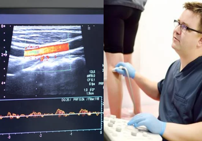
Private DVT ultrasound in Kent & London
Choose the clinic most convenient for you. The same focused DVT ultrasound assessment is available across all three locations.
Tunbridge Wells
Private DVT ultrasound of the leg or arm with compression and Doppler assessment, verbal feedback and same-day written reporting.
36 Monson Road
Tunbridge Wells, Kent
TN1 1LU
Orpington
Rapid-access private DVT ultrasound from our Orpington clinic, with focused deep venous assessment and same-day reporting.
Orpington Health & Wellbeing Centre
19 Homefield Rise
Orpington, BR6 0FE
Canary Wharf
Private DVT ultrasound in Canary Wharf with focused compression and Doppler assessment, clear explanation of findings and same-day written reporting.
30 S Colonnade
Canary Wharf Estate
London, E14 5HX
A focused ultrasound assessment of the deep veins
Deep Vein Thrombosis occurs when a blood clot forms in a deep vein, most commonly in the leg and less commonly in the arm. If part of a clot travels to the lungs, it can cause a pulmonary embolism.
DVT ultrasound is a non-invasive examination that combines compression of the veins with Doppler assessment of venous blood flow. It is used to assess whether a deep vein remains compressible and whether there are sonographic features of intraluminal clot. Where clinically relevant, the superficial venous system is also assessed for superficial venous thrombosis (superficial thrombophlebitis).
The examination is tailored to the symptomatic limb. When both limbs are selected, both sides are assessed during the same appointment.
Canary Wharf: £220 one limb · £400 both limbs
What does a DVT ultrasound examine?
The scan evaluates the deep venous system of the symptomatic limb using compression and Doppler techniques. Where clinically relevant, the superficial venous system is also assessed for superficial venous thrombosis / thrombophlebitis, with extension into relevant proximal or distal segments where indicated.
Deep venous system
Assessment of the principal deep veins in the symptomatic limb to look for sonographic evidence of deep venous thrombosis.
Superficial venous system
Where clinically relevant, superficial veins are assessed for superficial venous thrombosis (superficial thrombophlebitis), including the visible extent and relationship to the deep venous system.
Vein compressibility
Gentle probe pressure is used to assess whether the vein walls fully compress, an important part of the DVT examination.
Venous blood flow
Colour and pulsed Doppler are used where appropriate to assess venous flow and support interpretation of the examination.
Clot extent
When an abnormality is identified, the examination can be extended proximally or distally where clinically indicated to document its visible extent.
How the venous system is assessed
Simplified patient-education illustration of the deep venous pathway, with relevant superficial veins also assessed when clinically indicated.
Compression ultrasound and Doppler assessment
These complementary techniques assess both the physical compressibility of the vein and the pattern of blood flow within the deep venous system and relevant superficial veins.
Assessing vein compressibility
Ultrasound gel is applied to the skin and the probe is moved along the limb. Gentle pressure is applied at key deep and superficial venous segments, where relevant, to assess whether the vein compresses normally.
Assessing venous flow
Colour and pulsed Doppler can be used to assess blood flow within the veins, complementing compression findings and helping characterise the venous examination.
Symptoms and risk factors that may prompt DVT assessment
DVT ultrasound may be requested when symptoms or risk factors raise concern for a possible clot in a deep vein.
New limb swelling
New swelling of one leg or arm can be a reason for deep venous ultrasound assessment.
Pain or tenderness
New calf, thigh or limb pain and tenderness may require assessment where DVT is suspected.
Redness or warmth
Redness or warmth along the limb can occur alongside symptoms that prompt venous assessment.
Recent surgery or trauma
Recent surgery or significant trauma may increase concern when compatible limb symptoms develop.
Prolonged immobility
Reduced mobility, including prolonged travel, may be relevant when assessing the risk of DVT.
Previous DVT / PE or thrombophilia
A personal or family history of DVT, pulmonary embolism or thrombophilia may be clinically relevant.
Pregnancy
Pregnancy is one of the clinical contexts in which suspected DVT may require prompt venous ultrasound.
Hormonal therapy
Hormonal medication such as contraceptive therapy or HRT may be relevant to overall DVT risk.
Clinical follow-up
Some patients require ultrasound assessment because of a previous thrombotic history or ongoing clinical concern.
DVT requires a clear onward pathway when a clot is identified
The role of the ultrasound is to establish the venous imaging findings and support prompt onward clinical management.
DVT can lead to pulmonary embolism
A fragment of a deep venous clot can travel to the lungs. This is why prompt assessment and appropriate treatment are important when DVT is identified.
Positive findings require urgent clinical follow-up
If a clot is detected, the main findings are explained and you are guided toward urgent GP or appropriate specialist management, with a formal same-day report provided.
Ultrasound is part of the wider clinical pathway
Your ultrasound result should be interpreted together with symptoms, clinical history and any other medical assessment required by the treating clinician.
Superficial thrombophlebitis can also be identified
Where clinically relevant, the superficial veins are assessed for superficial venous thrombosis / thrombophlebitis and its visible relationship to the deep venous system.
What happens during your DVT ultrasound?
The examination is focused on the symptomatic limb and combines clinical history, deep venous compression ultrasound, assessment of relevant superficial veins, Doppler assessment and same-day reporting.
Clinical history
Your symptoms, relevant risk factors and previous DVT / PE history are reviewed before scanning.
Compression ultrasound
Gel is applied and the probe is used along the limb with gentle pressure to assess vein compressibility.
Doppler assessment
Colour and pulsed Doppler are used where indicated to evaluate venous blood flow.
Results & report
The main findings are explained and a formal written report is provided on the same day.
Preparing for your DVT ultrasound
No special preparation
You do not need to fast or change your normal eating and drinking routine for the DVT scan.
Wear loose clothing
Wear clothing that allows the Sonographer to access the relevant leg or arm comfortably.
Bring relevant information
Bring any relevant referral, recent test result or previous DVT / vascular imaging if available.
What Our Patients Say
Independent patient feedback from people who have chosen UKSONO Healthcare for private diagnostic ultrasound.
Guidance supporting DVT and venous ultrasound assessment
Selected UK guidance and patient information supporting the role of venous ultrasound in suspected DVT and superficial venous thrombosis.
Read NICE guidance →
Read SVT guidance →
Read NHS guidance →
DVT ultrasound FAQs
Answers to common questions about DVT ultrasound, compression testing, Doppler assessment, pricing and same-day results.
Do I need a GP referral?
How much does a DVT ultrasound cost?
How accurate is a DVT ultrasound?
How quickly will I receive the results?
Is the DVT scan painful?
How long does the scan take?
Do I need to prepare for the scan?
Can you scan an arm as well as a leg?
Can the scan assess superficial thrombophlebitis?
What happens if a blood clot is found?
Book your private DVT ultrasound
Focused deep venous ultrasound with compression and Doppler assessment, same-day reporting and direct online booking.