
Private Ultrasound & Health Checks in Canary Wharf.
Fast access to diagnostic ultrasound, pregnancy scans and advanced health checks at UNTIL Canary Wharf, 30 South Colonnade. Self-referral is available, with clear findings and reports usually provided on the same day.


Our specialist ultrasound and health services.
Choose the service that best matches your needs. Each option opens the relevant information page with preparation, pricing and booking guidance.
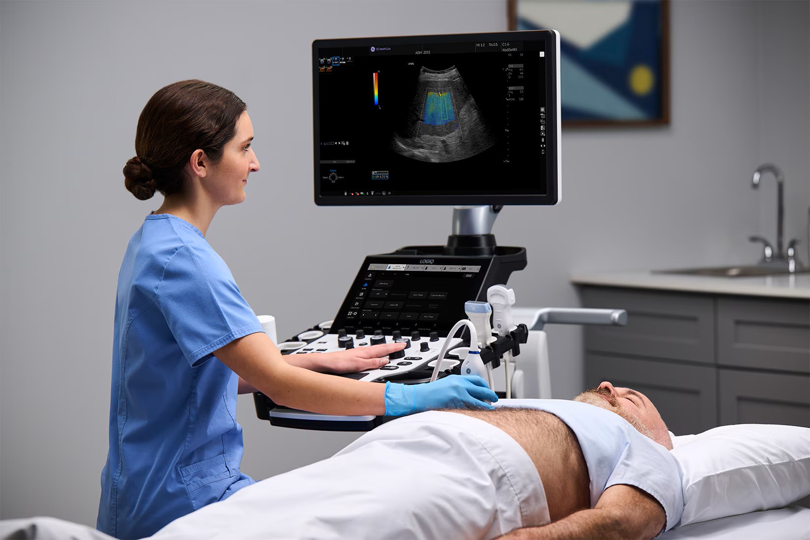 Advanced diagnostic ultrasound
Diagnostic Ultrasound Scans
Abdominal, pelvic, urinary, thyroid, neck, breast, testes, prostate, vascular and musculoskeletal ultrasound.
Explore diagnostic scans →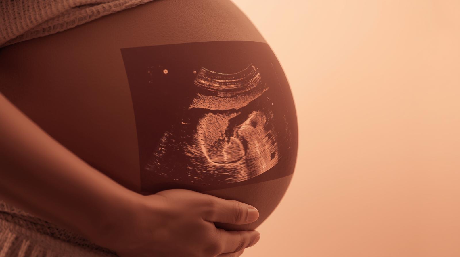 Pregnancy and fetal wellbeing
Pregnancy Ultrasound Scans
Early pregnancy, reassurance, dating, gender, anomaly, growth, Doppler and 3D/4D bonding scans.
Explore pregnancy scans → Men’s and family wellbeing
Men’s Health Check
A comprehensive men’s health assessment supporting long-term wellbeing, with ultrasound-led screening, targeted blood tests and clinical review options.
Explore men’s health checks → Women’s preventive health
Women’s Health Check
Comprehensive women’s health screening with ultrasound-led assessment, targeted blood tests and clinical review options.
Explore women’s health checks →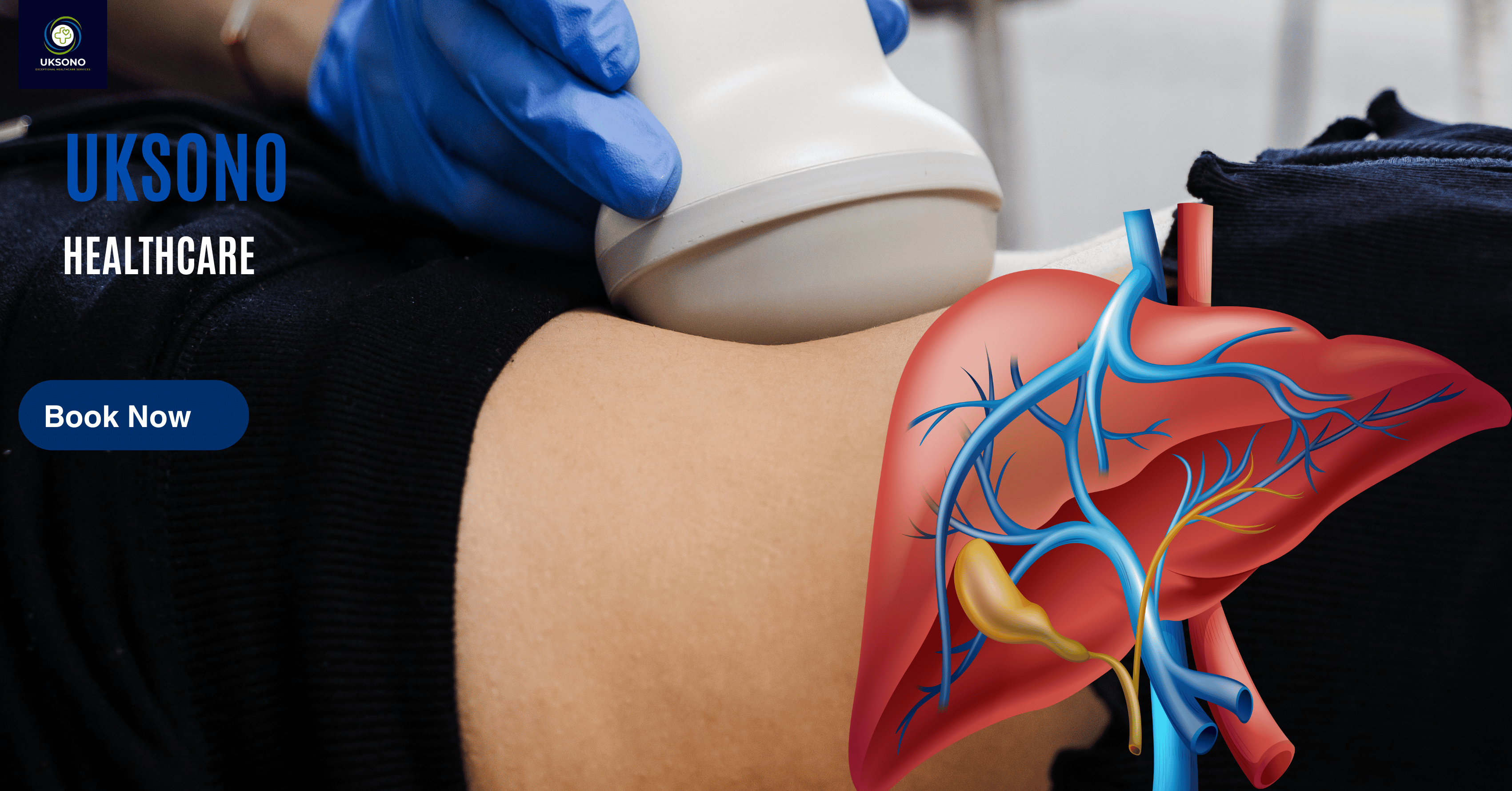 Liver structure, fat and fibrosis
Advanced Liver Health Checks
Liver ultrasound, fibrosis assessment, liver-fat measurement and selected blood-test pathways.
Explore liver checks → Thyroid structure and function
Thyroid Health Checks
Detailed thyroid and neck ultrasound with selected thyroid blood tests and onward guidance where appropriate.
Explore thyroid checks → Paediatric imaging · Ages 0–17
Paediatric Diagnostic Scans
Ages 0–17 yearsChild-focused diagnostic ultrasound for babies, children and young people aged 0–17 years, including suitable abdominal, urinary, neck and superficial soft-tissue concerns.
Explore paediatric scans →What scan do I need?
Describe your symptoms, body area, pregnancy stage or health concern in your own words. The finder can understand phrases and sentences and will display every closely related UKSONO service.
Frequently selected services
Type a concern or choose a category for tailored suggestions.
Try adding a body area or using a simpler symptom, or call 01892 251532 for guidance.
Browse all servicesClinical depth. London pace.
Designed for professionals, families and international visitors who value fast access without compromising clinical standards. Our Canary Wharf clinic combines advanced ultrasound, personalised health checks and clear communication in one considered patient journey.
Whether you need focused assessment of a symptom, pregnancy reassurance, a specialist liver or thyroid review, or a broader wellbeing check, our team helps you move from uncertainty to informed next steps.
Our London ultrasound clinic serves patients from Canary Wharf, Docklands, Tower Hamlets, Greenwich, the City of London and surrounding East London areas.
A clinical setting with presence.
Architectural calm, premium shared spaces and a private clinical environment designed to make healthcare feel considered from arrival to report.

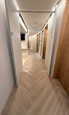

Canary Wharf clinic
A refined arrival in the heart of London’s business district.
Simple from start to finish.
Book directly
Select the scan or health check you need. A GP referral is not required.
Attend Canary Wharf
Arrive at 30 South Colonnade and check in within the premium UNTIL environment.
Understand your findings
Your sonographer explains the scan findings clearly, with appropriate next-step guidance.
Receive your report
Your written report and images are usually issued on the same day.
In the centre of Canary Wharf.
UKSONO Healthcare operates from the UNTIL health and wellness environment at YY London, 30 South Colonnade. The clinic is positioned for convenient access from Canary Wharf, Docklands, Tower Hamlets, East London and central London.
Trusted for clarity, care and professionalism.
Explore UKSONO Health Articles
Explore 50 uniquely written clinical guides combining patient-friendly explanations with dedicated epidemiology, pathology, diagnostic testing, ultrasound features, reporting points, differential diagnosis, management, prognosis and practical FAQs.
Liver Function Tests (LFTs): Parameters, Interpretation and Ultrasound Findings
ALT and AST mainly reflect hepatocellular injury; ALP and GGT point toward cholestasis or biliary involvement; bilirubin reflects haem breakdown and hepatic processing; albumin and INR provide more direct information about protein synthesis and clotting-factor production.
- Clinical background and anatomy
- Causes, symptoms and red flags
- Tests and common ultrasound findings
- Limitations, treatment and FAQs
Fatty Liver Disease: Causes, Symptoms, Ultrasound and Fibrosis Risk
Triglycerides accumulate within hepatocytes when lipid delivery and synthesis exceed oxidation and export. Some people remain at simple steatosis, while others develop steatohepatitis, fibrosis and eventually cirrhosis.
- Clinical background and anatomy
- Causes, symptoms and red flags
- Tests and common ultrasound findings
- Limitations, treatment and FAQs
Liver Fibrosis and Cirrhosis: Stages, Elastography and Ultrasound
Chronic injury activates stellate cells and extracellular-matrix deposition. Progressive fibrosis disrupts normal vascular and lobular architecture, increasing portal resistance and reducing functional reserve.
- Clinical background and anatomy
- Causes, symptoms and red flags
- Tests and common ultrasound findings
- Limitations, treatment and FAQs
Gallstones: Symptoms, Complications and Ultrasound Diagnosis
Cholesterol or pigment crystals form within bile and enlarge into stones. Symptoms typically arise when a stone obstructs the cystic duct; migration into the common bile duct can cause jaundice, cholangitis or pancreatitis.
- Clinical background and anatomy
- Causes, symptoms and red flags
- Tests and common ultrasound findings
- Limitations, treatment and FAQs
Gallbladder Polyps: Size, Risk and Ultrasound Follow-Up
Polyps represent mucosal projections. Common cholesterol polyps are non-neoplastic; adenomas and rare early carcinomas form the clinically important neoplastic group.
- Clinical background and anatomy
- Causes, symptoms and red flags
- Tests and common ultrasound findings
- Limitations, treatment and FAQs
Pancreatitis and Pancreatic Pain: Tests, Imaging and Red Flags
Premature pancreatic enzyme activation causes autodigestion and inflammation. Severe disease can lead to necrosis, fluid collections and systemic complications.
- Clinical background and anatomy
- Causes, symptoms and red flags
- Tests and common ultrasound findings
- Limitations, treatment and FAQs
Kidney Stones: Symptoms, Hydronephrosis and Ultrasound Findings
Crystals precipitate in supersaturated urine and aggregate into calculi. Obstruction occurs when a stone enters the ureter, increasing upstream pressure and causing colicky pain.
- Clinical background and anatomy
- Causes, symptoms and red flags
- Tests and common ultrasound findings
- Limitations, treatment and FAQs
Kidney Cysts: Simple, Complex and When Follow-Up Is Needed
Simple cysts arise from renal tubular structures and contain clear fluid. Complexity may reflect haemorrhage, infection or neoplastic tissue.
- Clinical background and anatomy
- Causes, symptoms and red flags
- Tests and common ultrasound findings
- Limitations, treatment and FAQs
Blood in the Urine: Causes, Ultrasound and Urology Assessment
Blood may originate anywhere from glomeruli to urethra. Structural, inflammatory, stone-related, prostatic and neoplastic causes must be considered.
- Clinical background and anatomy
- Causes, symptoms and red flags
- Tests and common ultrasound findings
- Limitations, treatment and FAQs
Enlarged Prostate: Symptoms, PSA, Ultrasound and Treatment
Hyperplasia of stromal and glandular tissue within the transition zone can narrow the urethra and increase bladder-outflow resistance.
- Clinical background and anatomy
- Causes, symptoms and red flags
- Tests and common ultrasound findings
- Limitations, treatment and FAQs
Testicular Lump: Common Causes and Ultrasound Assessment
Lumps may arise from the testis, epididymis, spermatic cord, hydrocele sac, skin or hernia. Determining anatomical origin is central to diagnosis.
- Clinical background and anatomy
- Causes, symptoms and red flags
- Tests and common ultrasound findings
- Limitations, treatment and FAQs
Scrotal Pain: Torsion, Infection, Varicocele and Ultrasound
Pain may result from vascular compromise, infection, inflammation, trauma, hernia, varicocele or referred pain.
- Clinical background and anatomy
- Causes, symptoms and red flags
- Tests and common ultrasound findings
- Limitations, treatment and FAQs
Breast Lump: Benign Causes, Red Flags and Ultrasound
A palpable lesion may arise from glandular tissue, ducts, fat, skin or inflammatory change. The key clinical task is to distinguish benign from suspicious pathology.
- Clinical background and anatomy
- Causes, symptoms and red flags
- Tests and common ultrasound findings
- Limitations, treatment and FAQs
Breast Cysts: Symptoms, Ultrasound Features and Management
Fluid accumulates within terminal duct-lobular units. Cysts may be simple, complicated by internal debris or complex with true solid components.
- Clinical background and anatomy
- Causes, symptoms and red flags
- Tests and common ultrasound findings
- Limitations, treatment and FAQs
Armpit Lump (Axillary Lump): Causes, Symptoms and Ultrasound
The axilla contains lymph nodes, fat, skin appendages, vessels and accessory breast tissue. Pathology may arise from any of these structures.
- Clinical background and anatomy
- Causes, symptoms and red flags
- Tests and common ultrasound findings
- Limitations, treatment and FAQs
Neck Lump: Causes, Clinical Assessment and Ultrasound
A neck mass may arise from lymph nodes, thyroid, salivary glands, congenital remnants, skin or other soft tissues.
- Clinical background and anatomy
- Causes, symptoms and red flags
- Tests and common ultrasound findings
- Limitations, treatment and FAQs
Thyroid Nodules: Ultrasound Risk Features and Next Steps
Nodules include colloid hyperplasia, cysts, adenomas, inflammatory nodules and a small minority of thyroid cancers.
- Clinical background and anatomy
- Causes, symptoms and red flags
- Tests and common ultrasound findings
- Limitations, treatment and FAQs
Goitre: Causes, Thyroid Function and Ultrasound Assessment
Diffuse or nodular enlargement may reflect increased stimulation, inflammation or clonal nodule formation.
- Clinical background and anatomy
- Causes, symptoms and red flags
- Tests and common ultrasound findings
- Limitations, treatment and FAQs
Swollen Lymph Nodes: Reactive Change, Red Flags and Ultrasound
Nodes enlarge through immune-cell proliferation, inflammation, infiltration by malignancy or infection.
- Clinical background and anatomy
- Causes, symptoms and red flags
- Tests and common ultrasound findings
- Limitations, treatment and FAQs
Hernias: Types, Symptoms and Dynamic Ultrasound Diagnosis
Fat, bowel or other tissue protrudes through an anatomical defect or weakened fascial layer.
- Clinical background and anatomy
- Causes, symptoms and red flags
- Tests and common ultrasound findings
- Limitations, treatment and FAQs
Groin Lump: Hernia, Lymph Nodes and Other Causes
The lump may arise from the inguinal canal, femoral canal, lymph nodes, skin, fat or vessels.
- Clinical background and anatomy
- Causes, symptoms and red flags
- Tests and common ultrasound findings
- Limitations, treatment and FAQs
Abdominal Pain: Which Organs Can Ultrasound Assess?
Pain may arise from solid organs, bowel, urinary tract, vessels, abdominal wall or referred thoracic and musculoskeletal sources.
- Clinical background and anatomy
- Causes, symptoms and red flags
- Tests and common ultrasound findings
- Limitations, treatment and FAQs
Right Upper Quadrant Pain: Gallbladder, Liver and Other Causes
Pain may arise from gallbladder obstruction/inflammation, liver capsule distension, right kidney, stomach/duodenum, lung base or abdominal wall.
- Clinical background and anatomy
- Causes, symptoms and red flags
- Tests and common ultrasound findings
- Limitations, treatment and FAQs
Pelvic Pain: Gynaecological Causes and Ultrasound Assessment
Pain may originate from uterus, ovaries, tubes, bladder, bowel, pelvic floor or musculoskeletal structures.
- Clinical background and anatomy
- Causes, symptoms and red flags
- Tests and common ultrasound findings
- Limitations, treatment and FAQs
Ovarian Cysts: Types, Ultrasound Features and Follow-Up
Cysts may be follicular, corpus-luteal, haemorrhagic, endometriotic, dermoid, benign neoplastic or malignant.
- Clinical background and anatomy
- Causes, symptoms and red flags
- Tests and common ultrasound findings
- Limitations, treatment and FAQs
Fibroids: Types, Symptoms, Fertility and Ultrasound Mapping
Fibroids are monoclonal smooth-muscle tumours whose growth is influenced by ovarian hormones and genetic factors.
- Clinical background and anatomy
- Causes, symptoms and red flags
- Tests and common ultrasound findings
- Limitations, treatment and FAQs
Endometriosis: Symptoms, Ultrasound and Diagnostic Limitations
Endometrial-like tissue outside the uterine cavity induces cyclical bleeding, inflammation, fibrosis and adhesions.
- Clinical background and anatomy
- Causes, symptoms and red flags
- Tests and common ultrasound findings
- Limitations, treatment and FAQs
Adenomyosis: Symptoms, Ultrasound Features and Treatment
Endometrial glands and stroma within the myometrium induce muscular hypertrophy and architectural distortion.
- Clinical background and anatomy
- Causes, symptoms and red flags
- Tests and common ultrasound findings
- Limitations, treatment and FAQs
PCOS: Hormones, Ultrasound Criteria and Fertility
The condition involves dysregulation of ovarian androgen production, follicular development and insulin signalling. Polycystic ovarian morphology is only one component.
- Clinical background and anatomy
- Causes, symptoms and red flags
- Tests and common ultrasound findings
- Limitations, treatment and FAQs
Female Infertility Assessment: Ultrasound, AMH and Hormones
Successful conception requires ovulation, adequate ovarian reserve, tubal patency, normal uterine environment and viable sperm.
- Clinical background and anatomy
- Causes, symptoms and red flags
- Tests and common ultrasound findings
- Limitations, treatment and FAQs
Follicular Tracking: Ovulation Monitoring and Scan Timing
A cohort of follicles develops each cycle; one typically becomes dominant and grows until ovulation while the endometrium responds to oestrogen and progesterone.
- Clinical background and anatomy
- Causes, symptoms and red flags
- Tests and common ultrasound findings
- Limitations, treatment and FAQs
Bleeding in Early Pregnancy: Causes, Ultrasound and Red Flags
Bleeding may arise from implantation-site change, subchorionic haemorrhage, cervical disease, pregnancy failure or extrauterine implantation.
- Clinical background and anatomy
- Causes, symptoms and red flags
- Tests and common ultrasound findings
- Limitations, treatment and FAQs
Miscarriage: Symptoms, Ultrasound Diagnosis and Follow-Up
Pregnancy development arrests and trophoblastic support eventually fails, producing bleeding and uterine evacuation or retained products.
- Clinical background and anatomy
- Causes, symptoms and red flags
- Tests and common ultrasound findings
- Limitations, treatment and FAQs
Ectopic Pregnancy: Warning Signs, hCG and Ultrasound
Implantation occurs outside the endometrial cavity, most commonly within the fallopian tube. Tubal rupture can cause life-threatening internal bleeding.
- Clinical background and anatomy
- Causes, symptoms and red flags
- Tests and common ultrasound findings
- Limitations, treatment and FAQs
Deep Vein Thrombosis (DVT): Symptoms and Duplex Ultrasound
Venous stasis, endothelial injury and hypercoagulability promote thrombus formation within deep veins.
- Clinical background and anatomy
- Causes, symptoms and red flags
- Tests and common ultrasound findings
- Limitations, treatment and FAQs
Varicose Veins and Venous Insufficiency: Doppler Mapping
Failure of venous valves allows retrograde flow, increasing ambulatory venous pressure and producing varicosities, oedema and skin change.
- Clinical background and anatomy
- Causes, symptoms and red flags
- Tests and common ultrasound findings
- Limitations, treatment and FAQs
Carotid Artery Disease: Plaque, Stenosis and Stroke Risk
Atherosclerotic plaque develops within the arterial wall and may narrow the lumen or embolise material to cerebral arteries.
- Clinical background and anatomy
- Causes, symptoms and red flags
- Tests and common ultrasound findings
- Limitations, treatment and FAQs
Abdominal Aortic Aneurysm: Risk Factors and Ultrasound Screening
Degeneration of the aortic media and extracellular matrix leads to permanent arterial dilatation.
- Clinical background and anatomy
- Causes, symptoms and red flags
- Tests and common ultrasound findings
- Limitations, treatment and FAQs
Shoulder Pain: Rotator Cuff, Bursitis and Ultrasound
Pain can arise from tendon degeneration or tearing, bursal inflammation, calcific deposits, impingement or joint pathology.
- Clinical background and anatomy
- Causes, symptoms and red flags
- Tests and common ultrasound findings
- Limitations, treatment and FAQs
Tennis Elbow: Tendon Changes, Ultrasound and Treatment
The common extensor tendon develops degenerative collagen change and microtearing rather than simple acute inflammation.
- Clinical background and anatomy
- Causes, symptoms and red flags
- Tests and common ultrasound findings
- Limitations, treatment and FAQs
Wrist Ganglion: Symptoms, Ultrasound and Treatment Options
A mucin-filled cyst arises from a joint capsule or tendon sheath and may communicate with the underlying joint.
- Clinical background and anatomy
- Causes, symptoms and red flags
- Tests and common ultrasound findings
- Limitations, treatment and FAQs
Plantar Fasciitis: Heel Pain, Ultrasound and Recovery
Repetitive tensile load causes degenerative change at the calcaneal origin of the plantar fascia.
- Clinical background and anatomy
- Causes, symptoms and red flags
- Tests and common ultrasound findings
- Limitations, treatment and FAQs
Achilles Tendon Injury: Tendinopathy, Tears and Ultrasound
Tendinopathy reflects collagen disorganisation and failed tendon adaptation. Rupture occurs when tensile load exceeds tendon capacity.
- Clinical background and anatomy
- Causes, symptoms and red flags
- Tests and common ultrasound findings
- Limitations, treatment and FAQs
Baker’s Cyst: Knee Swelling, Ultrasound and Management
Fluid distends the gastrocnemius-semimembranosus bursa and may communicate with the knee joint.
- Clinical background and anatomy
- Causes, symptoms and red flags
- Tests and common ultrasound findings
- Limitations, treatment and FAQs
Thyroid Function Tests: TSH, Free T4, T3 and Antibodies
The hypothalamic-pituitary-thyroid axis regulates TSH and thyroid hormones through negative feedback.
- Clinical background and anatomy
- Causes, symptoms and red flags
- Tests and common ultrasound findings
- Limitations, treatment and FAQs
Full Body Ultrasound: Benefits, Limitations and Incidental Findings
There is no single disease mechanism because multiple organs are assessed. The clinical value comes from identifying structural abnormalities accessible to ultrasound.
- Clinical background and anatomy
- Causes, symptoms and red flags
- Tests and common ultrasound findings
- Limitations, treatment and FAQs
Women’s Health Checks: Ultrasound, Blood Tests and Screening
A comprehensive check may consider reproductive, endocrine, cardiovascular, metabolic and structural health rather than treating these as isolated systems.
- Clinical background and anatomy
- Causes, symptoms and red flags
- Tests and common ultrasound findings
- Limitations, treatment and FAQs
Men’s Health Checks: Prostate, Testes, Blood Tests and Risk
A comprehensive check integrates metabolic risk with structural assessment rather than using ultrasound as a stand-alone test.
- Clinical background and anatomy
- Causes, symptoms and red flags
- Tests and common ultrasound findings
- Limitations, treatment and FAQs
Liver Health Checks: Ultrasound, FibroScan and Blood Tests
Different tests assess different dimensions: blood tests reflect injury and function, ultrasound assesses structure, attenuation assesses fat and elastography estimates stiffness.
- Clinical background and anatomy
- Causes, symptoms and red flags
- Tests and common ultrasound findings
- Limitations, treatment and FAQs
Which Ultrasound Scan Do I Need? A Patient Decision Guide
Different body systems require different imaging modalities because ultrasound penetration, field of view and tissue characteristics vary.
- Clinical background and anatomy
- Causes, symptoms and red flags
- Tests and common ultrasound findings
- Limitations, treatment and FAQs
No matching article found
Try a broader symptom, organ or condition.
Before you arrive.
Do I need a GP referral?
No. UKSONO Healthcare accepts self-referrals for private ultrasound scans and health checks.
How quickly will I receive my report?
Ultrasound reports are usually issued on the same day. Blood-test turnaround depends on the selected package and laboratory testing requirements.
Where is the Canary Wharf clinic?
The clinic is within UNTIL at YY London, 30 South Colonnade, Canary Wharf Estate, London E14 5HX.
Which scans are available?
Services include diagnostic abdominal, pelvic, urinary, liver, thyroid, neck, breast, testes, prostate, vascular and musculoskeletal ultrasound, alongside pregnancy scans and advanced health-check packages. Availability may vary by appointment date.
Clarity, in the heart of London.
Book your private ultrasound scan or health assessment directly. No GP referral is required.
Same-day clarity. Specialist care. Zero compromise.
At UKSONO Healthcare, our mission is to deliver fast, accurate and patient-centred diagnostic ultrasound and health checks. We pair senior clinical expertise with state-of-the-art technology to provide clear answers, empower proactive decisions, and bridge the gap between NHS delays and private care — with compassion, clarity and trust.