
Fatty Liver Disease (MASLD)
A detailed guide to metabolic dysfunction-associated steatotic liver disease: what causes fat to accumulate in the liver, who is at risk, how blood tests and ultrasound contribute to diagnosis, how fibrosis is assessed and what you can do to reduce long-term liver and cardiovascular risk.
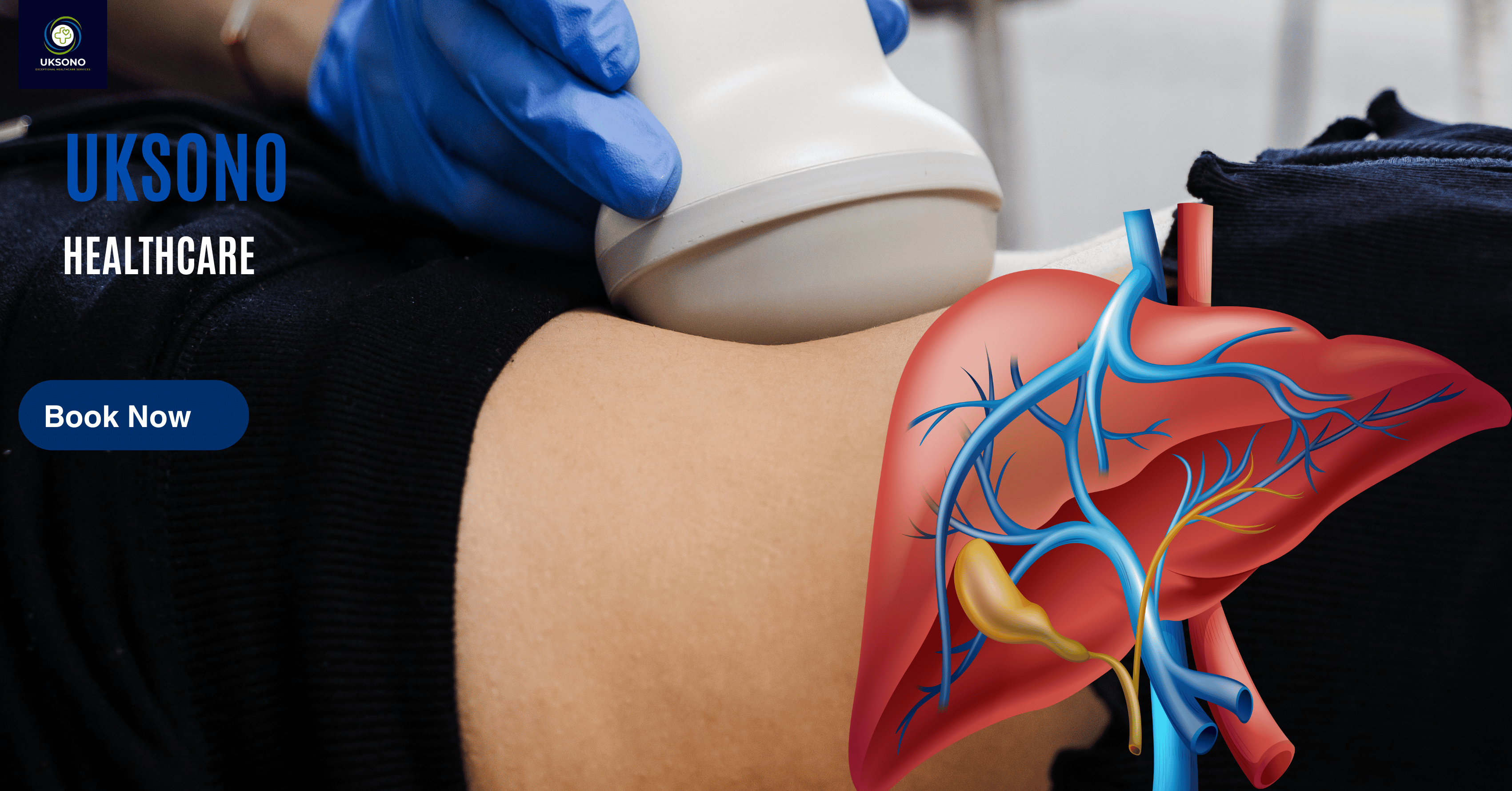
Overview: What is fatty liver disease?
MASLD sits within a broader group of conditions called steatotic liver disease. Some people have fat in the liver without meaningful inflammation or scarring. Others develop metabolic dysfunction-associated steatohepatitis (MASH), where steatosis is accompanied by hepatocellular injury and inflammation. Persistent injury can lead to fibrosis, then cirrhosis in a smaller proportion of patients.
The condition is closely linked to cardiometabolic health. Obesity, increased abdominal or visceral fat, insulin resistance, type 2 diabetes, dyslipidaemia and high blood pressure are important associations. This is why modern management focuses not only on the liver but also on cardiovascular, metabolic and kidney risk.
Fatty liver is frequently discovered incidentally. A routine abdominal ultrasound may show a bright or echogenic liver, or blood tests may reveal an abnormal ALT, AST or GGT. Importantly, routine liver blood tests can be normal even when steatosis or clinically significant fibrosis is present. NICE specifically advises against using routine liver blood tests alone to rule out fatty liver disease or advanced fibrosis.
What does the liver actually do?
The liver is one of the body’s major metabolic organs. It sits predominantly in the right upper abdomen beneath the diaphragm and receives blood from both the hepatic artery and portal vein. Its functions are wide-ranging, which is why significant chronic liver disease can affect many systems.
Metabolism
The liver helps regulate glucose, cholesterol, triglycerides and amino acids. It stores glycogen and releases glucose when needed. It also converts and packages fats for transport.
Protein production
It produces albumin and many clotting factors. Falling albumin or a prolonged clotting time can be a sign of impaired synthetic function in advanced disease, although these usually remain normal in early MASLD.
Bile production
Bile made by the liver helps digest and absorb dietary fats. It travels through the intrahepatic and extrahepatic bile ducts before reaching the bowel.
Detoxification & processing
The liver metabolises many medicines, hormones and toxins and helps process bilirubin produced during red blood cell breakdown.
Why is it called MASLD rather than NAFLD?
The terminology for fatty liver disease has changed. The older term non-alcoholic fatty liver disease (NAFLD) defined the condition largely by what it was not: significant alcohol-related disease. The newer terminology focuses more directly on the metabolic drivers of disease.
| Older terminology | Current terminology | What it means |
|---|---|---|
| NAFLD | MASLD | Steatotic liver disease in a person with one or more cardiometabolic risk factors, without another dominant explanation such as harmful alcohol intake. |
| NASH | MASH | Metabolic dysfunction-associated steatohepatitis: steatosis with hepatocellular injury and inflammation, which can promote fibrosis. |
| Alcohol-related / mixed categories | MetALD and ALD | Current nomenclature recognises that metabolic dysfunction and alcohol exposure can coexist rather than forcing every patient into a single binary category. |
You will still see NAFLD and NASH in older NICE guidance, research papers, clinic letters and imaging reports. The underlying clinical concepts overlap substantially, so a previous diagnosis of NAFLD does not suddenly become invalid because the terminology has changed.
How common is fatty liver disease?
MASLD is one of the most common chronic liver disorders worldwide. It is particularly common in people with type 2 diabetes, obesity and metabolic syndrome. NICE notes that NAFLD is present in more than half of adults with type 2 diabetes or metabolic syndrome in the evidence underpinning its research recommendations.
Not everyone with liver fat develops advanced disease. The clinical challenge is identifying the smaller group with progressive inflammation and fibrosis. This is why modern pathways increasingly use staged risk assessment rather than treating every bright liver on ultrasound as equivalent.
A more useful question is: “Do I have evidence that the liver is being injured or scarred, and what cardiometabolic factors are driving that risk?”
What causes MASLD?
MASLD develops when energy and lipid handling become dysregulated and excess fat accumulates in hepatocytes. Insulin resistance plays a central role. Increased delivery of fatty acids from adipose tissue, increased production of fat within the liver and altered export or oxidation of fat all contribute.
Common risk factors
- Overweight or obesity, especially increased abdominal or visceral fat.
- Type 2 diabetes or insulin resistance.
- High triglycerides or other dyslipidaemia.
- High blood pressure.
- Metabolic syndrome.
- Polycystic ovary syndrome (PCOS), particularly where insulin resistance is present.
- Obstructive sleep apnoea and other conditions associated with metabolic dysfunction.
- Low physical activity and diets high in ultra-processed foods or sugar-sweetened drinks.
- Family/genetic susceptibility.
Can lean people develop MASLD?
Yes. MASLD is not restricted to people with obesity. The 2024 European guideline recognises normal-weight MASLD. A person can have a BMI within the normal range but still have increased visceral adiposity, insulin resistance, reduced muscle mass or genetic susceptibility. For that reason, body weight is an important risk marker but not the whole story.
Weight is important, but MASLD is a metabolic condition influenced by fat distribution, insulin sensitivity, genetics, diet, activity, age and other cardiometabolic factors.
What about alcohol?
Alcohol history remains important. Significant alcohol exposure can itself cause steatosis and progressive liver disease, and alcohol can add to metabolic liver risk. The newer nomenclature recognises overlap categories such as MetALD. Patients should give clinicians an accurate alcohol history rather than assuming that a diagnosis of “fatty liver” automatically means alcohol is irrelevant.
What are the symptoms of fatty liver disease?
Most people with early MASLD have no specific symptoms. The NHS notes that many people are diagnosed only because blood tests or scans were performed for another reason.
Possible early symptoms
- Persistent tiredness or low energy.
- A general sense of being unwell.
- Right upper abdominal discomfort beneath the ribs.
- Symptoms related to associated diabetes or metabolic disease rather than the liver itself.
Symptoms of advanced disease
- Abdominal swelling from ascites.
- Leg swelling.
- Jaundice.
- Easy bruising or bleeding.
- Muscle loss or weight loss.
- Confusion or altered concentration in decompensated disease.
A person can have clinically important fibrosis while feeling well. Conversely, fatigue or abdominal discomfort can have many causes unrelated to the liver.
Stages of fatty liver disease
Fatty liver disease exists on a spectrum. Progression is not inevitable, and patients may move in either direction depending on metabolic control, weight, alcohol exposure, treatment and individual susceptibility.
Why fibrosis matters
Liver fat can fluctuate relatively quickly with metabolic changes. Fibrosis reflects a longer-term wound-healing response to persistent injury. Advanced fibrosis is a stronger predictor of liver-related outcomes than steatosis alone, so fibrosis assessment is an important part of modern MASLD care.
How is MASLD diagnosed?
There is no single test that answers every question. Diagnosis usually combines the clinical history, cardiometabolic risk profile, alcohol assessment, liver blood tests and imaging. Additional blood tests may be used to exclude alternative causes of liver disease.
A typical diagnostic pathway
- Identify steatosis or risk: abnormal ultrasound, abnormal liver blood tests or significant cardiometabolic risk.
- Review alternative causes: alcohol, medicines, viral hepatitis, autoimmune disease, haemochromatosis and other relevant conditions depending on the presentation.
- Assess fibrosis risk: blood-based scores such as FIB-4 or other locally used tests.
- Use second-line fibrosis testing where indicated: transient elastography, shear-wave elastography, ARFI-based techniques or ELF depending on local pathways.
- Refer higher-risk patients: hepatology assessment when advanced fibrosis, cirrhosis, diagnostic uncertainty or another concerning feature is present.
The 2024 EASL–EASD–EASO guideline supports case-finding for liver fibrosis in people with cardiometabolic risk factors, abnormal liver enzymes and/or radiological steatosis, with particular attention to type 2 diabetes and obesity with additional metabolic risk factors. A staged approach using FIB-4 followed by imaging-based elastography is specifically highlighted.
Liver blood tests explained
“Liver function tests” is a convenient phrase, but not every component directly measures liver function. Some markers reflect hepatocellular injury, others cholestasis, and others synthetic capacity. Interpretation depends on the pattern, degree of abnormality and clinical context.
| Test | What it broadly reflects | How it can relate to MASLD |
|---|---|---|
| ALT | Hepatocellular injury | May be raised in MASH or other liver injury, but can be normal despite steatosis or fibrosis. |
| AST | Hepatocellular injury; also found outside the liver | Patterns and ratios may change as disease advances, but AST is not specific to MASLD. |
| ALP | Cholestatic/biliary processes; also bone | A marked isolated rise should prompt consideration of biliary or non-hepatic causes rather than being attributed automatically to fatty liver. |
| GGT | Enzyme induction / cholestatic pattern | Can be raised in metabolic liver disease and with alcohol or medicines; it is not specific. |
| Bilirubin | Bilirubin production, hepatic processing and excretion | Usually normal in uncomplicated early MASLD. Jaundice requires investigation. |
| Albumin | Hepatic protein synthesis, but affected by other conditions | Often normal until advanced liver disease; low albumin has a broad differential diagnosis. |
| INR / prothrombin time | Clotting-factor synthesis | Prolongation can signal reduced hepatic synthetic reserve in advanced disease, although anticoagulants and other factors must be considered. |
| Platelets | Not a liver enzyme; useful in fibrosis risk scores | A falling platelet count can be seen with portal hypertension and is incorporated into FIB-4. |
NICE advises that routine liver blood tests should not be used to rule out fatty liver disease or advanced fibrosis. Imaging and fibrosis risk assessment may still be appropriate when the clinical risk is significant.
What is FIB-4?
FIB-4 is a simple fibrosis risk score derived from age, AST, ALT and platelet count. It is useful as a first-stage risk stratification tool rather than as a definitive diagnosis. Thresholds require age-aware interpretation and local clinical pathways. Intermediate or high-risk results often prompt a second-line test such as elastography or ELF.
What does fatty liver look like on ultrasound?
Conventional B-mode ultrasound is widely used because it is non-invasive, accessible, does not use ionising radiation and can assess the liver together with the gallbladder, bile ducts, kidneys, spleen and other upper abdominal structures.
Typical ultrasound features of hepatic steatosis
- Increased liver echogenicity: the liver appears brighter than expected, often compared with the right renal cortex when the kidney is suitable for comparison.
- Reduced visualisation of intrahepatic vessels: vessel walls and deeper vascular detail may become less conspicuous.
- Posterior beam attenuation: ultrasound penetration decreases with depth, making the deep liver and diaphragm harder to see.
- Reduced diaphragm definition: in more marked steatosis the posterior liver-diaphragm interface may be less distinct.
- Hepatomegaly: some patients have an enlarged liver, although size alone is not diagnostic of steatosis.
Conventional ultrasound can identify or suggest steatosis and assess morphology, but it cannot reliably determine whether microscopic steatohepatitis is present. That distinction relates to hepatocellular injury and inflammation, historically established with histology when tissue diagnosis is required.
Can ultrasound grade mild, moderate and severe fatty liver?
Sonographers may describe the visual severity of steatosis, but conventional qualitative grading has limitations. It is affected by machine settings, body habitus, fibrosis, operator technique and the subjective nature of comparing echogenicity. Modern quantitative attenuation techniques aim to provide a more reproducible estimate of liver fat.
Quantitative ultrasound fat assessment
Newer ultrasound platforms can estimate how strongly the liver attenuates the ultrasound beam. Because fat changes acoustic attenuation, these measurements can provide an objective parameter related to steatosis rather than relying only on a visual “bright liver” impression.
UGAP and attenuation-based techniques
Ultrasound-guided attenuation parameter (UGAP) and similar vendor-specific methods measure attenuation within a defined liver region of interest. The result is usually expressed in attenuation units such as dB/cm/MHz. Thresholds vary by technology and validation study, so results must be interpreted using the correct manufacturer and evidence base rather than applying one machine’s cut-off to another.
Quantitative fat assessment can be useful for baseline characterisation and longitudinal monitoring, particularly when performed on the same platform under similar conditions. It does not by itself tell you the fibrosis stage or prove MASH.
Elastography, liver stiffness and FibroScan
Elastography estimates tissue stiffness. In chronic liver disease, fibrosis tends to increase liver stiffness, which makes elastography useful for non-invasive fibrosis risk assessment.
Shear-wave elastography (SWE)
Shear-wave elastography uses ultrasound-generated shear waves and measures their propagation through the liver. Results may be displayed in metres per second (m/s) or converted to kilopascals (kPa). The examination is performed using defined quality criteria, typically through an intercostal window with a region of interest placed in appropriate liver parenchyma away from large vessels and artefacts.
FibroScan / vibration-controlled transient elastography
FibroScan uses vibration-controlled transient elastography to estimate liver stiffness. NICE HealthTech guidance recognises FibroScan as a non-invasive method for assessing fibrosis and cirrhosis in defined pathways. FibroScan can also provide a controlled attenuation parameter (CAP) to estimate steatosis.
Liver stiffness is influenced by fibrosis but can also rise with active inflammation, cholestasis, hepatic congestion, recent food intake and technical factors. Cut-offs vary by disease population and technology. The result should be interpreted with the clinical context and quality metrics.
Why use both fat and stiffness measurements?
They answer different questions. Attenuation-based measures estimate the amount of steatosis; elastography estimates stiffness and therefore fibrosis risk. A patient can have substantial fat with little fibrosis, or less obvious fat but clinically important fibrosis after years of disease.
CT, MRI and liver biopsy: when are they used?
| Test | Strengths | Important limitations |
|---|---|---|
| Ultrasound | Accessible, no ionising radiation, assesses liver morphology and other abdominal organs; can be combined with Doppler, attenuation imaging and elastography. | Mild steatosis may be difficult to detect; qualitative grading is subjective; image quality can be limited by body habitus and bowel gas. |
| CT | Can demonstrate steatosis and is excellent for many complications or alternative abdominal diagnoses. | Uses ionising radiation and is not usually performed simply to screen for uncomplicated fatty liver. |
| MRI / MRI-PDFF | Highly accurate non-invasive quantification of liver fat; MR elastography can assess fibrosis. | More expensive, less accessible, and not required for most routine cases. |
| Liver biopsy | Can directly assess steatosis, inflammation, ballooning and fibrosis and remains important in selected diagnostic dilemmas. | Invasive, carries procedural risk and sampling variability; unsuitable as a population screening test. |
Cross-sectional imaging or biopsy is generally reserved for situations where it will answer a clinically important question not adequately resolved by routine non-invasive assessment.
How is fatty liver disease treated?
Treatment is personalised according to fibrosis risk, body weight, diabetes status, cardiovascular risk, alcohol intake and other comorbidities. There is no single universal “fatty liver cure.” The foundation is reducing the metabolic drivers of liver injury.
1. Improve diet quality
The 2024 European MASLD guideline recommends improving diet quality in a pattern similar to a Mediterranean diet, limiting ultra-processed foods rich in sugar and saturated fat, and avoiding sugar-sweetened beverages. The practical goal is a sustainable eating pattern rather than a short restrictive “liver detox.”
2. Exercise regularly
Physical activity can reduce liver fat and improve insulin sensitivity even when weight loss is modest. The NHS advises aiming for at least 150 minutes of exercise each week for people with fatty liver disease, adapted to individual health and capability.
3. Manage diabetes, lipids and blood pressure
Cardiovascular disease is a major source of morbidity in MASLD. Optimal management of type 2 diabetes, hypertension and dyslipidaemia is therefore central to treatment, not an optional extra.
4. Address alcohol intake
Alcohol can compound liver injury. Patients with MASLD should discuss alcohol exposure with their healthcare professional, particularly if fibrosis is present. NHS advice for the general population remains not to exceed 14 units per week, but a lower or zero intake may be appropriate for some people with liver disease.
5. Stop smoking
Smoking increases cardiovascular and cancer risk and undermines overall metabolic health. Stopping smoking is one of the most valuable long-term health interventions a person can make.
How much weight loss improves fatty liver?
When a person with MASLD is overweight, the magnitude of sustained weight loss matters. The 2024 EASL–EASD–EASO guideline gives useful evidence-based targets:
Weight loss should be safe, sustainable and individualised. Rapid crash diets can be difficult to maintain and may be unsuitable for some patients. People with advanced liver disease, frailty, eating disorders, pregnancy or major medical conditions should seek personalised advice before undertaking a significant weight-loss plan.
European guidance recommends diet and exercise interventions even in normal-weight adults with MASLD because liver fat can improve without obesity being present.
Are there medicines for MASLD or MASH?
Pharmacological treatment is evolving rapidly. Medicines may be prescribed primarily for obesity, diabetes, cardiovascular risk or selected liver indications. Treatment should be clinician-led rather than based on a scan result alone.
GLP-1 receptor agonists and incretin-based therapy
Medicines such as semaglutide and tirzepatide may be indicated for type 2 diabetes or obesity in appropriate patients and can produce substantial weight loss and metabolic improvement. European guidance supports their use for their licensed metabolic indications in people with MASLD, while distinguishing this from recommending every GLP-1 drug as a direct MASH-targeted therapy.
Resmetirom
Resmetirom has been approved in some jurisdictions for selected adults with non-cirrhotic MASH and significant fibrosis. Availability and licensing vary by country, and treatment decisions require specialist assessment. It should not be viewed as a general treatment for everyone with an ultrasound diagnosis of fatty liver.
Pioglitazone, vitamin E and other treatments
Older guidelines and selected specialist pathways discuss pioglitazone or vitamin E for particular groups. Evidence and recommendations depend on diabetes status, fibrosis stage, age and setting. These should not be self-started for fatty liver.
“Natural” does not mean liver-safe. Herbal and dietary supplements can themselves cause liver injury or interact with prescribed medication. Discuss supplements with a pharmacist, GP or liver specialist.
Monitoring, follow-up and prognosis
Monitoring should be driven by fibrosis risk and metabolic health rather than repeating a conventional ultrasound at arbitrary intervals. Follow-up may include weight, blood pressure, HbA1c/glucose, lipid profile, liver blood tests, fibrosis scores and repeat non-invasive fibrosis assessment.
NICE recommends assessment for advanced liver fibrosis in people diagnosed with NAFLD. Its current NG49 pathway uses the ELF test, with specialist referral for advanced fibrosis. In real-world UK practice, availability varies, and FibroScan or other elastography techniques are frequently used alongside blood-based risk scores.
What determines prognosis?
- Fibrosis stage.
- Presence and control of type 2 diabetes.
- Obesity and visceral adiposity.
- Cardiovascular disease and overall metabolic risk.
- Alcohol exposure.
- Age and other liver diseases.
- Whether lifestyle and metabolic risk factors improve over time.
Early steatosis can improve substantially. Advanced fibrosis and cirrhosis require closer specialist follow-up because risks include portal hypertension, liver decompensation and hepatocellular carcinoma. NICE recommends six-monthly liver ultrasound surveillance for hepatocellular carcinoma in people with cirrhosis without hepatitis B, with or without serum AFP according to the pathway.
When should you seek medical help?
Arrange routine medical review if:
- An ultrasound reports hepatic steatosis or features of chronic liver disease.
- Liver enzymes remain abnormal on repeat testing.
- You have type 2 diabetes, obesity or metabolic syndrome and are concerned about liver risk.
- You have unexplained persistent right upper abdominal discomfort or fatigue.
- A fibrosis score or elastography result is abnormal or indeterminate.
New jaundice, vomiting blood, black stools, marked abdominal swelling, confusion, collapse, severe persistent abdominal pain or rapidly worsening illness should not wait for a routine private ultrasound appointment.
Frequently asked questions
Can fatty liver be seen on ultrasound?
Yes. Moderate or marked hepatic steatosis often produces characteristic increased echogenicity and attenuation. Mild steatosis may be less conspicuous, and image quality can be affected by body habitus and other technical factors.
Can I have fatty liver with normal ALT and AST?
Yes. Normal routine liver enzymes do not exclude steatosis or fibrosis. NICE specifically advises against using routine liver blood tests alone to rule out fatty liver disease or advanced fibrosis.
Does fatty liver always progress to cirrhosis?
No. Many people remain at the steatosis stage, particularly when metabolic risk factors are improved. A smaller group develops progressive inflammation and fibrosis. Fibrosis risk assessment helps identify people who need closer follow-up.
Can fatty liver be reversed?
Liver fat can fall substantially with sustained improvements in weight, diet, physical activity and metabolic control. MASH can also improve. Fibrosis can regress in some people, but advanced fibrosis is more difficult to reverse, which is why early intervention matters.
What is the difference between fatty liver and fibrosis?
Steatosis describes excess fat within liver cells. Fibrosis is scar tissue produced after persistent injury. A person can have significant steatosis with little fibrosis, or advanced fibrosis after years of liver injury even when current fat levels are less striking.
Does a conventional liver ultrasound tell me my fibrosis stage?
Not reliably. Advanced chronic liver disease may produce morphological signs on B-mode ultrasound, but early fibrosis cannot be staged accurately from brightness or texture alone. Elastography and/or validated blood-based tests are used for non-invasive fibrosis risk assessment.
What is the difference between FibroScan and shear-wave elastography?
Both are non-invasive elastography methods used to estimate liver stiffness. FibroScan uses vibration-controlled transient elastography, whereas shear-wave elastography is integrated into many diagnostic ultrasound systems. They have different acquisition methods and validated cut-offs, so results are not interchangeable without context.
Is fatty liver caused by eating fat?
The biology is more complex. MASLD reflects disordered energy metabolism and insulin resistance, influenced by calorie excess, visceral adiposity, dietary composition, genetics and physical activity. Simply removing all dietary fat is not the recommended treatment.
Should I stop alcohol completely?
This depends on your liver status and overall risk. Alcohol can add to metabolic liver injury. People with fibrosis or cirrhosis should discuss alcohol specifically with their clinician; abstinence may be advised.
How often should liver elastography be repeated?
There is no single interval appropriate for every person. Repeat testing depends on baseline fibrosis risk, diabetes and metabolic control, changes in weight, abnormal blood tests and specialist recommendations.
How UKSONO Healthcare can help
A useful liver assessment should answer more than “is the liver bright?” Depending on the selected UKSONO liver-health package and clinical indication, assessment can combine diagnostic liver ultrasound with advanced non-invasive methods for liver fat and fibrosis risk, plus selected blood testing.
Structural assessment
- Liver morphology and echotexture.
- Gallbladder and biliary tract.
- Portal and hepatic venous appearances where relevant.
- Spleen, ascites and features that may suggest portal hypertension.
- Focal liver lesions visible on ultrasound.
Advanced liver assessment
- Quantitative attenuation / liver-fat assessment where included.
- Shear-wave elastography / stiffness assessment where included.
- FibroScan-type pathways depending on package/location.
- Selected blood tests and metabolic risk assessment.
- Clear recommendations for GP or hepatology follow-up when indicated.
Concerned about fatty liver or liver fibrosis?
Explore UKSONO Healthcare’s advanced liver health checks for assessment options, preparation guidance, locations and current package details.
Explore Advanced Liver Health Checks Tunbridge Wells · Orpington · Canary Wharf · 01892 251532References & further reading
- European Association for the Study of the Liver (EASL), European Association for the Study of Diabetes (EASD), European Association for the Study of Obesity (EASO). EASL–EASD–EASO Clinical Practice Guidelines on the management of metabolic dysfunction-associated steatotic liver disease (MASLD). Journal of Hepatology. 2024;81(3):492–542. View guideline.
- NICE. Non-alcoholic fatty liver disease (NAFLD): assessment and management (NG49). Published 2016; reviewed 2024. View NICE guidance.
- NICE. Cirrhosis in over 16s: assessment and management (NG50). Last updated September 2023. View NICE guidance.
- NICE. FibroScan for assessing liver fibrosis and cirrhosis outside secondary and specialist care (HTG682). 2023. View NICE HealthTech guidance.
- NHS. Non-alcoholic fatty liver disease (NAFLD). View NHS patient information.
- NICE. MRI-based technologies for assessing non-alcoholic fatty liver disease (HTG655). 2023. View NICE HealthTech guidance.
The terminology and evidence base for steatotic liver disease are evolving. UKSONO articles should be periodically reviewed against updated NICE, NHS, EASL, BSG and other relevant specialty guidance. References are provided so readers and clinicians can verify key recommendations.